Outline of “A case study which critically evaluates the role of the diagnostic imaging in the management and treatment of a patient presenting with a renal lesion”

Introduction

60-year-old patient presented to her GP with haematuria and a persistent cough. Urine and blood samples may have  indicated issues with serum creatinine and glomerular filtration rate (European Association of Urology, 2017). The patient had reduced liver function which led to biologic treatment for tuberculosis being discontinued one year previously. In keeping with recommendations (NHS, 2016),  patient was referred to the urology department from the GP for an ultrasound. This essay considers the contribution of diagnostic imaging to the patient’s management in terms of diagnosis, staging, treatment and followup. 

Diagnosis

-Ultrasound

DoH targets (2011) -patients with suspected renal malignancy to be diagnosed within 2 weeks

-Ultrasound is quick, relatively cheap and accessible

 differentiates between solid and cystic masses, indicates size and vascularity (Rossi et al., 2018). Authors are multi-disciplinary UK professionals, making source particularly relevant to this patient’s care. However, some citations are old therefore some information may not be up to date. However, other sources have supported the accuracy of ultrasound, particularly with contrast enhancement, to characterize kidney lesions

sens: 88%, spec: 80%- Wang et al., 2014; sens: 96%, spec: 50%- Chang et al., 2017.

The patient’s examination identified a 6cm kidney lesion.

Staging

-CT

sensitivity: 60%, spec: 73%, PPV: 43%, NPV: 84%- Millet et al. (2011) 

The patient underwent a CT scan of the chest, abdomen and pelvis with contrast. “gold standard choice” (Rossi et al., 2018).This is in accordance with recommendations of a contrast-enhanced CT scan of the abdomen and thorax for staging following the identification of a suspicious lesion. In addition, MR imaging or a bone scan is suggested (NHS England, 2016).

-MRI

Despite MRI being available, patient did not have an MRI scan potentially due to the waiting time or contraindications.

-Nuclear medicine bone scan

This demonstrated multiple areas of increased uptake.  Following these scans,  patient was discussed at MDT in accordance with NHS guidance (2016). The case is complicated by known latent tuberculosis infection. Since myobacteria is repressed by the patient’s immune system, undergoing immunocompromising cancer treatment could have reactivate the TB (Nachiappan et al., 2017).  Thus team were keen to ascertain whether the lesion was infectious or malignant as this greatly affected the treatment plan.

-Ultrasound-guided vs CT biopsy

Treatment (NICE, 2019)

Interventional procedure– percutaneous or laparoscopic nephrectomy, radiofrequency ablation or cryotherapy 

Drugs– Cabozantinib, Tivozanib, Pazopanib, Sunitinib 


Follow up

Diagnostic imaging to assess the success of treatment

How well managed the tuberculosis infection is 

CT

FDG-PET/CT sens: 46.6%, spec: 66.6% –Olzuker et al. (2011)

References 

Capitanio, U. and Montorsi, F. (2016) Renal Cancer. The Lancet [online]. 387(10021), pp. 894-906.

Department of Health and Social Care (2011) Improving outcomes: a strategy for cancer [online]. London: Department of Health. (15108). Available from: https://assets.publishing.service.gov.uk/government/uploads/system/uploads/attachment_data/file/213785/dh_123394.pdf [Accessed 1 March 2019].

European Association of Urology (2017) EAU Guidelines on Renal Cell Carcinoma: The 2017 Update [online]. Available from: uroweb.org/wp-content/uploads/10-Renal-Cell-Carcinoma_2017_web.pdf [Accessed 28 February 2019]. 

Millet,  I., Doyon, F., Hoa, D., Thuret, R., Merigeaud, S., Serre, I. and Taourel, P. (2011) Characterization of Small Solid Renal Lesions: Can Benign and Malignant Tumors Be Differentiated With CT? AJR [online]. 197(4), pp. 887-896.  [Accessed 1 March 2019].

National Health Service England (2016) Guidelines for the Management of Renal Cancer (online). Available from: https://www.england.nhs.uk/mids-east/wp-content/uploads/sites/7/2018/05/guidelines-for-the-management-of-renal-cancer.pdf [Accessed 25 February 2019]. 

Nachiappan, A., Rahbar, K., Shi, X., Guy, E., Barbosa, E., Shroff, G., Ocazionez, D., Schlesinger, A., Katz, S. and Hammer, M. (2017) Pulmonary Tuberculosis: Role of Radiology in Diagnosis and Management. RSNA (online). 37 (1), pp. 52-72. [Accessed 27 February 2019]. 

National Institute for Health and Care Excellence (2015) Suspected cancer: recognition and referral [online]. London: Department of Health. (NG12). Available from: https://www.nice.org.uk/guidance/ng12/evidence/full-guideline-pdf-2676000277 [Accessed 25 February 2019].  

National Institute for Health and Care Excellence (2019) Renal Cancer Overview Flowchart [online]. London: Department of Health. Available from: https://pathways.nice.org.uk/pathways/renal-cancer#content=view-node%3Anodes-procedures-for-treating-renal-cancer [Accessed 25 February 2019].

Ozulker T, Ozulker F, Ozbek E, (2011) A prospective diagnostic accuracy study of F-18 fluorodeoxyglucose-positron emission tomography/computed tomography in the evaluation of indeterminate renal masses. Nuclear Medicine Communications [online]. 32 (no issue number), pp.265–272. 

Rossi, S., Prezzi, D., Kelly-Morland, C. and Goh, V. (2018) Imaging for the diagnosis and response assessment of renal tumoursWorld Journal of Urology [online]. 36(12), pp.1927-1944.  

Wang, C., Yu, C., Yang, F., Yang, G. (2014) Diagnostic accuracy of contrast-enhanced ultrasound for renal cell carcinoma: a meta-analysis. Tumor Biology [online]. 35(7), pp. 6343–6350.  

Wang, H., Ding, H., Chen, J., Chao, C., Meta-analysis of the diagnostic performance of [18F]FDG-PET and PET/CT in renal cell carcinoma. Cancer Imaging [online]. 12(3), pp. 464-474.


Which modality is ideal for imaging Crohn’s Disease?

It is thought that one in every 650 people in the UK has been diagnosed with Crohn’s disease, a chronic, inflammatory disease of the gastrointestinal tract which evidences itself through pain, diarrhoea, anaemia and weight loss. It occurs most commonly in the ileum, jejunum or colon, but can also present in the oesophagus, stomach or duodenum. There is increased incidence of the disease in women, smokers, and certain ethnic groups. Patients are typically diagnosed between the ages of 10 and 40 (Crohn’s and Colitis UK, 2013), with up to a third receiving a diagnosis before the age of 21 (Gomez, 2000).  Interestingly, there has previously been thought to be an increased incidence in twins proposing a genetic cause (Tysk et al., 1988). However, more recent studies have suggested that this study overestimated the influence of genetics on Crohn’s Disease (Halfvarson, 2011). Sufferers typically experience acute symptomatic attacks followed by periods of remission. Complications, such as abscesses, intestinal obstruction, fistulae, strictures and perforation, can arise and additionally, Crohn’s disease is also associated with joint, liver, skin and eye abnormalities (National Institute for Health and Care Excellence (NICE), 2012). As a result, patients who suffer from Crohn’s disease are likely to undergo a great deal of imaging in the management of their condition. Because of the nature of the disease, images are required to demonstrate not only anatomical indicators, such as mucosal ulcers, stones, separated bowel loops and thickened folds, but also provide functional information about activity. There are a vast number of examinations which can be used to image Crohn’s disease and its complications. This post considers some of those which can be used in the evaluation of Crohn’s disease, as well as the factors and circumstances which must be taken into account when making a decision.

According to UK patient-facing information, the initial investigation of patients presenting with Crohn’s disease-like symptoms involves a physical examination, a blood test for anaemia and a stool test for signs of bleeding and inflammation (Crohn’s and Colitis UK, 2013; National Health Service, 2018). Prior to deciding the method of disease management which could be drug therapy, nutritional changes or surgery, imaging is needed to determine the location, extent and severity of the disease.

Abdomen x-rays are accessible to GPS for referral and can be used to demonstrate kidney stones, obstruction and perforation, yet they cannot provide information on the location of inflammation, thickness of the bowel wall or the disease’s effect on vasculature. There is more of a role for x-ray in in following-up complications. Because Crohn’s disease is a secondary cause of osteoporosis, the NICE guidelines recommend dual energy x-ray absorptiometry (DEXA) to monitor bone density in young people identified with risk factors such as low body mass index (BMI) and glucocorticosteroid use (NICE, 2012). Plain film imaging may also be required as patients get older in order to diagnose or monitor arthritis.

Traditionally for diagnosis, barium follow-through films were used but cross-sectional imaging is increasingly replacing barium fluoroscopy in small bowel imaging as it can assess location, extent, severity and complications simultaneously (Taylor et al., 2018). The use of contrast is valuable in assessing wall thickness and patterns. Yet, Ilangovan et al. (2012) suggest that using barium can conceal intraluminal haemorrhage and so should not be used unless it is needed to highlight an obstruction. However, this seems counterproductive as administration of large amounts of contrast is not advisable when querying an obstruction because in the event of a positive diagnosis, it could exacerbate the problem. Wireless capsule endoscopy is an interesting, relatively new and painless option for patients in whom a gastrointestinal bleed is queried. It involves swallowing a capsule containing a camera which images the patient’s bowel as it travels through over the course of ten hours (NICE, 2004; Cotter et al., 2014).

More invasive procedures include colonoscopy, and endoscopy which can be time-consuming , require sedation, a nursing team and a hospital bed for recovery afterwards. Thus, they are far from ideal but, for example, are still recommended by NICE guidelines to screening patients who have experienced Crohn’s disease symptoms for more than 10 years for colonic cancer (2011). Endoscopic surveillance is recommended for evaluating remission in patients following surgery (NICE, 2012).

CT

CT enteroclysis involves the insertion of a nasojejunal tube for the administration of water as a neutral contrast material in addition to an injection of intravenous contrast (Gianlunca et al., 2012). Ilangovan et al. (2012) argue that, while a neutral contrast provides maximum contrast between the lumen and bowel wall, water will provoke rapid reabsorption and thus result in suboptimal distension. Good distension and contrast enhancement demonstrates abnormal bowel loops well, an indication of mesenteric fat changes, as well as highlighting any fistulae or ulceration. Despite producing optimal contrast enhancement, enteroclysis is an unpleasant procedure for the patient and entails the risk of perforation. An alternative is enterography which requires the patient to drink oral contrast over 40-60 minutes rather than be intubated, which is more tolerable to patients but requires their compliance (Ilangovan et al., 2012). This could be an issue with paediatric patients. A drawback of both these procedures is that they are time-consuming and incur a radiation dose of which the average is 15mSv (Ilangovan et al, 2012). This could be further reduced through use of automated exposure controls, tube axes modulation and adjusting the parameters and exposure factors. For an older Crohn’s disease sufferer, in whom radiation dose is not such a concern, CT is perhaps the most relatively inexpensive, informative and accessible option. It is important to consider, however, the mean sensitivity and specificity of CT in 33 studies was calculated as 84.3% and 95.1% respectively (Horsthuis et al., 2008). While still high, the sensitivity is lower than ultrasound, MRI and nuclear medicine. Consequently, depending on the accuracy required, another modality may be more suitable.

MRI

Kim and Ha (2003) describe magnetic resonance (MR) enteroclysis as the most accurate method of visualising Crohn’s disease, since it provides good distension of bowel loops. A major benefit of magnetic resonance imaging (MRI) is the ability to produce high quality, multiplanar images with excellent soft tissue contrast without radiation exposure. Yet, as the study does not mention which type of scanner was used in the research, it may not be comparable to more modern technology.  Hence it may be a little outdated, and lacks acknowledgement of more recent developments, such as 3D rendering, virtual colonoscopy, and ‘fly-through’ which can be additionally valuable in bowel assessment. However, this conclusion is supported by Gianluca et al.’s more recent review which maintains its validity (2012). Taylor et al. (2018) highlight that MRI is indeed highly accurate with a sensitivity of 97% for detecting disease presence and 80% for extent of disease.  The use of a large sample size of 284 patients from 8 different UK hospitals supports the study’s reliability, generalisability and relevance to UK healthcare practice. In practice, enterography is preferred over enteroclysis, likely due to the lack of intubation needed, despite producing inferior bowel distension (Gianluca et al., 2012).  Lymph node enhancement and contrast uptake by the small bowel wall demonstrates the extent of disease activity (Kim and Ha, 2003). Scanning in different sequences provides visualisation of the mesenteric vasculature which is important as activity is indicated by enlargement and engorgement of these. This produces a so-called “comb sign” which is demonstrated by Figure 1 (Ilangovan, 2012).  Considering that patients with Crohn’s disease require follow-up imaging and often receive their diagnosis while young, it is important to limit radiation exposure as much as possible, therefore in addition to its high sensitivity, MRI appears an ideal option (Panes et al., 2011).

comb sign

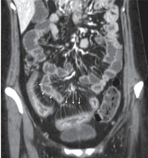
Figure 1: A CT scan demonstrating the “comb sign” From British Journal of Radiology (2012)

Pregnancy care

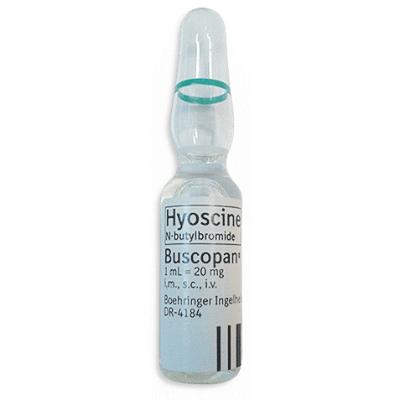
Stern el al. (2014) studied the effectiveness of an MR enterography protocol in pregnancy. While its international location (Israel) and use of a small sample size (n=9) may affect the generalisability of this research to the UK, it remains useful in highlighting MRE as a valuable tool in the investigation of Crohn’s in pregnant women while minimising foetal risks. Crohn’s disease carries the risks of pre-term delivery and low birth weight hence the importance of imaging, not only to check foetal growth, but also to monitor the mother’s symptoms. Computed tomography is generally a less favoured modality in pregnancy due its risk of radiation dose to the foetus. While literature demonstrates a lack of consensus over potential acoustic, heating and magnetic field effects on the foetus, MRI is considered to have no known adverse effects (Bulas and Egloff, 2013). Despite this, in the UK it is advised that a scan should be postponed until after delivery if possible, and that if a scan must go ahead, gradient echo sequences and acoustic lessening modes should be utilised to reduce any potential adverse effects. The greatest issue is the use of gadolinium-based contrast which is not recommended in pregnancy unless absolutely necessary due to concerns over potential deposits left in the body (Society of Radiographers, 2016). Mannitol was utilised in this study instead, while fast-acquisition mode can compensate for the lack of glucagon or Buscopan, minimising the effect of bowel movement on image quality (Stern et al. 2014).

While seemingly the best modality to use, in reality MRI may not be accessible to a patient due to location or the length of waiting times and it is a comparatively expensive modality. Contraindications, such as some pacemakers or metal foreign bodies, as well as claustrophobia, may also prevent it from being a realistic modality option for patients with a chronic condition. For pregnant patients, there may be image interpretation difficulties as soft tissue differentiation is lessened, in addition to anatomical changes which have taken place in the abdomen naturally due to pregnancy. Therefore, it may necessary to use a combination of MRI and ultrasound to increase accuracy.

Ultrasound

Considering its relative low cost, accessibility, lack of radiation and patient discomfort, ultrasound is a valuable modality for monitoring, especially in the care of paediatrics and pregnant women. It allows the measurement of wall thickening, demonstrates narrowing of the lumen, and confirms the presence of fistulae, perienteric fat, increased lymph nodes and abscesses, with the highest sensitivity in more accessible areas such as the ileum and left colon (Gianluca et al, 2012). There are also the added benefits of using Doppler to analyse the superior mesenteric artery flow volume as well as the interventional potential of endoscopic ultrasound to take a biopsy for histological examination. The study by Taylor et al. (2018) found that in detecting the presence of Crohn’s disease ultrasound has a good sensitivity of 92% and a specificity of 84%. Therefore, it is accurate enough to be a valuable imaging tool in the initial investigation of Crohn’s disease. Because it also has sensitivity of 70% and specificity of 81% in measuring the extent of disease, it can also be used in follow-up investigations. Although MRI is clearly much more accurate in the measurement of extent (sensitivity=80%; specificity=95%), it is not as accessible or as cheap as ultrasound. However, ultrasound’s accuracy is somewhat dependent upon the operator, patient size, and the location of the disease.

Nuclear medicine

99m Tc-HMPAO-labelled Leukocyte Scintigraphy (TLLS) is theoretically an ideal technique to use since it highlights the areas of inflammation throughout the whole body, impacting on the next stage of the patient pathway. As it shows arthritis, it highlights not only the severity of the disease in the gastrointestinal tract, but also complications of it. Rispo et al. (2005) found that the sensitivity when combining this technique and ultrasound was 100%. While this study had a decent sample size of 80 patients, only 50 were diagnosed with Crohn’s disease which may affect its validity. Yet, its statistical results were comparable to the slightly more recent study by Horsthuis et al. (2008) which used a much larger sample size from 33 studies (Table 1). Gianluca et al. (2012) describe a number of advantages including the lack of risks or contraindications and good levels of toleration, however examinations are time consuming and are best for highlighting acute cases due to low anatomical resolution. Also, there is a requirement for specialist staff training which may mean that this modality is inaccessible to some patients.

Table 1: Comparison of research studies’ results of the accuracy of different modalities and examinations in the assessment of Crohn’s Disease

Modality Sensitivity (%) Specificity (%) Reference About the Research
Ultrasound Conventional ultrasound 83-87 95-99 Panes et al. (2011) Systematic review of studies into Crohn’s disease imaging. Selected 5 studies including 1029 patients for analysis- large sample size
92 84 Taylor et al. (2018) A UK multicentre trial published in a reputable, peer-reviewed journal The Lancet.

Large sample size of 284 and the fact that multiple centres were used promotes its reliability, generalisability and relevance

87.8 84.5 Horsthuis et al. (2008) Published in Radiology- endorsed by Radiological Society of North America. Large sample size using data from 33 studies. Limitations are that sensitivity and specificity values are means so accuracy is affected. Also, the research is generalised to include inflammatory bowel diseases rather than just Crohn’s disease
92.0 97.0 Rispo et al. (2005) Published in peer-reviewed journal Inflammatory Bowel Diseases

Sample size of 80 patients; 50 had Crohn’s disease, therefore the results may relate more generally to abdominal imaging rather than Crohn’s disease specifically

90.3 93.7 Migaleddu et al. (2009) Published in reputable journal (American Gastroenterological Association). Image evaluation was by 2 blinded radiologists.

Limitations include small sample size (47 patients) however these are restricted only to patients with Crohn’s Disease

Contrast enhanced ultrasound 93.5 93.7 Migaleddu et al. (2009) As above.
Doppler 90.3 93.7 Migaleddu et al. (2009) As above.
Nuclear Medicine 99mTc-HMPAO 90.0 93.0 Rispo et al. (2005) As above.
89.7 95.6 Horsthuis et al. (2008) As above.
MRI Enteroclysis


93.0 92.8 Horsthuis et al. (2008) As Above.
78.0 85.0 Panes et al. (2011) As above.
  Enterography 97.0 96 Taylor et al. (2018) As above.
CT   84.3 95.1 Horsthuis et al (2008) As above.


Conclusion

There is not a single ‘gold standard’ method of imaging Crohn’s disease (Gianluca et al., 2012), as patients with Crohn’s disease have a very complex healthcare pathway. Choices must reflect the chronic nature of the condition and thus minimising the radiation dose should be a priority. Procedures should be low cost, non-invasive and well-tolerated. As the majority of Crohn’s sufferers will undergo surgery at some point, they may require imaging during theatre and for complications which require them to undergo other examinations such as DEXA or CT scans. Therefore, in terms of long-term management of Crohn’s disease, ideally MRI and ultrasound should be used to keep the radiation dose of young patients to a minimum. Research has shown that both modalities can accurately confirm the presence of the disease, while MRE has the highest sensitivity in monitoring the extent and severity of the disease. Compared with optical imaging modalities, it provides high quality cross sectional analysis of bowel structure with reduced patient discomfort, as well as information about the surrounding blood vessels, which is important in forming a treatment plan.  However, there are issues with accessibility, waiting times, contraindications and cost which may mean that CT is used instead. The continued development of hybrid imaging, especially PET/MRI, may be valuable in the assessment of Crohn’s Disease since it would produce highly accurate anatomical information with functional information about activity. This could be another interesting blog topic.


References: 

Bulas, D and Egloff, A. (2013) Benefits and risks of MRI in pregnancy. Seminars in Perinatology [online]. 37(5), pp. 301-304. [Accessed 28 November 2018]. 

Cotter, J., Dias de Castro, F., Moreira, M. and Rosa, B. (2014) Tailoring Crohn’s disease treatment: The impact of small bowel capsule endoscopy. Journal of Crohn’s and Colitis [online]. 8(12), pp. 1610-1615. [Accessed 28 November 2018]. 

Crohn’s and Colitis UK (2013) Crohn’s Disease [online]. London: Crohn’s and Colitis UK. Available from: https://www.crohnsandcolitis.org.uk/about-inflammatory-bowel-disease/crohns-disease [Accessed 23 November 2018]. 

Gomez J. (2000) Living with Crohn’s Disease (Overcoming Common Problems). London: Sheldon Press. 

Gianluca, G., Graziella, G., Veronica, M., Cinzia, L., Luigi, M., Ilario, S., Antonio, R. and Roberto, G. (2012) Crohn’s Disease Imaging: A Review. Gastroenterology Research and Practice [online]. No volume (no issue number), pp.1-15. [Accessed 23 November 2018]. 

Halfvarson (2011) Genetics in twins with Crohn’s disease: Less pronounced than previously believed? Inflammatory Bowel Diseases [online]. 17(1), pp.6-12. [Accessed 26 November 2018]. 

Horsthuis, K., Bipat, S., Bennink, R. and Stoker, J. (2008) Inflammatory Bowel Disease Diagnosed with US, MR, Scintigraphy, and CT: Meta-analysis of Prospective Studies. Radiology [online]. 247(1). [Accessed 28 November 2018]. 

Ilangovan, R., Burling, D., George, A., Gupta, A., Marshall, M. and Taylor, S. (2012) CT enterography: review of technique and practical tips. British Journal of Radiology [online]. 85(no issue number), pp. 876-886. [Accessed 24 November 2018]. 

Kim, K. and Ha, H. (2003) MRI for small bowel diseases. Seminars in Ultrasound, CT and MRI [online]. 24(5), pp. 387-402. [Accessed 14 November 2018]. 

Migaleddu, V., Scanu, A., Quaia, E., Rocca, P., Dore, M., Scanu, D., Azzali, L. and Virgilio, G. (2009) Contrast-enhanced ultrasonographic evaluation of inflammatory activity in Crohn’s disease. Gastroenterology [online]. 137(1), pp. 43-52. [Accessed 28 November 2018]. 

National Health Service (2018) Crohn’s Disease. Available from: https://www.nhs.uk/conditions/crohns-disease/diagnosis [Accessed 23 November 2018]. 

National Institute for Health and Care Excellence (NICE) (2004) Wireless capsule endoscopy for investigation of the small bowel [online]. London: Department of Health. (IPG101). Available from: https://www.nice.org.uk/guidance/ipg101 [Accessed 28 November 2018]. 

National Institute for Health and Care Excellence (NICE) (2011) Colorectal cancer prevention: colonoscopic surveillance in adults with ulcerative colitis, Crohn’s disease or adenomas [online]. London: Department of Health. (CG118). Available from: https://www.nice.org.uk/guidance/cg118 [Accessed 23 November 2018]. 

National Institute for Health and Care Excellence (NICE) (2012) Crohn’s Disease: Management in adults, children and young people[online]. London: Department of Health. (CG152). Available from: https://www.nice.org.uk/guidance/cg152 [Accessed 23 November 2018]. 

Panes, J., Bouzas, R., Chaparro, M., Garcia-Sanchez, V., Gisbert, J., Martinez de Guerenu, B., Mendoza, J., Paredes, J., Quiroga, S., Ripolles, T. and Rimola, J. (2011) Systematic review: the use of ultrasonography, computed tomography and magnetic resonance imaging for the diagnosis, assessment of activity and abdominal complications of Crohn’s disease. Alimentary Pharmacology and Therapeutics [online]. 34(2), pp. 125-145. [Accessed 23 November 2018]. 

Paredes, J., Ripolles, T., Cortes, X., Reyes, M., Lopez, A., Martinez, M. and Moreno-Ossett, E. (2010) Non-invasive diagnosis and grading of postsurgical endoscopic recurrence in Crohn’s disease: Usefulness of abdominal ultrasonography and 99m Tc-hexamethylpropylene amineoxime-labelled leucocyte scintigraphy. Journal of Crohn’s and Colitis [online]. 4(1), pp.537-545. [Accessed 23 November 2018]. 

Rispo, A., Imbriaco, M., Celentano, L., Cozzolino, A., Camera, L., Mainenti, P., Manguso, F., Sabbatini, F., D’Amico, P. and Castiglione, P. (2005) Inflammatory Bowel Diseases [online]. 11(4), pp. 376-382. [Accessed 28 November 2018]. 

Society of Radiographers (2016) Safety in Magnetic Resonance Imaging [online]. London: Society of Radiography. Available from: https://www.sor.org/learning/document-library/safety-magnetic-resonance-imaging-1 [Accessed 29 November 2018].

Stern, M., Kopylov, U., Ben-Horin, S., Apter, S. and Amitai, M. (2014) Magnetic resonance enterography in pregnant women with Crohn’s disease: case series and literature review. BMC Gastroenterology [online]. 14(no issue number), pp. 1-9. [Accessed 24 November 2018]. 

Taylor, S., Mallett, S., Bhatnagar, G., Baldwin-Cleland, R., Bloom, S., Gupta, A., Hamlin, P., Hart, A., Higginson, A, Jacobs, I., McCartney, S., Miles, A., Murray, C., Plum, A., Pollok, R., Punwani, S., Quinn, L., Rodriguez-Justo, M., Shabir, Z., Slater, A., Tolan, D., Travis, S., Windsor, A, Wylie, P., Zealley, I. and Halligan, S. (2018) Diagnostic accuracy of magnetic resonance enterography and small bowel ultrasound for the extent and activity of newly diagnosed and relapsed Crohn’s disease (METRIC): a multicentre trial. The Lancet [online]. 3(8), pp. 548-558. [Accessed 26 November 2018]. 

Tysk, C., Lindberg, E., Jarnerot, G., Floderus-Myrhed, B. (1988) Ulcerative colitis and Crohn’s disease in an unselected population of monozygotic and dizygotic twins. A study of heritability and the influence of smoking. BMJ [online]. 29(7), pp. 990-996. [Accessed 26 November 2018]. 
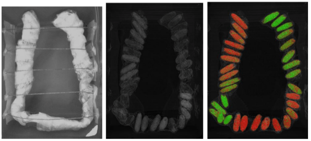
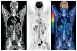
Buscopan: Cause for Concern?

Hyosine butylbromide, better known as Buscopan, relaxes the smooth muscle of the abdominal and pelvic cavities and is therefore recommended to aid image quality and reduce patient discomfort during several radiological examinations, including colonoscopy, endoscopy and prostate cancer detection (Lee et al., 2018; Barentisz et al., 2012). A UK survey found that 85.6% of gastroenterologists regularly use Buscopan in colonoscopy (Bedford et al., 2012). This post explores the benefits and risks of administering Buscopan, including recent safety alerts.

Buscopan ampoule for injection – from kaysmedical.com

The British Society of Gastroenterology advises that Buscopan helps to reduce the effect of peristaltic contractions on gastrointestinal imaging, increasing lesion detection by 30% (Lee et al., 2018). Despite this, a study by Nagata et al. (2015) disputes the routine use of Buscopan for computed tomography colonography, concluding that it does not clinically affect colonic distension, and creates unnecessary adverse effects and costs. The study compares colonic distension and patient tolerance of Buscopan administration with CO2 sufflation using a sample size of 208. The results showed that colonic distension increased marginally by a mean of just 0.06 compared to the control sample when using Buscopan. However, potential limitations of the study include the use of a 4-point grading system to measure colonic distension, three of which were classified as suboptimal and only one optimal, which suggests that the results are somewhat subjective and possibly biased. The assumption is made that all patients adhered to the pre-scan preparation instructions which, if they did not, may have affected the results. Image quality was not measured as part of the study, which is an issue since the Royal College of Radiographers (2017) conclude that the danger of poor image quality and subsequent misdiagnosis of cancer is of greater danger to patients than any adverse effects. The assessment of patient tolerance is challenging since it carries the potential to be subjective, also the questionnaire did not specifically address patient discomfort. Using the mean for result analysis eliminates patients for whom Buscopan may have created a much more positive experience. Therefore, in the UK, use of Buscopan alongside CO2 sufflation for colongraphy remains the routine procedure (Cancer Research UK, 2016).

Administration of Buscopan has known effects on the parasympathetic nervous system, increasing the heart rate and decreasing blood pressure (RCR, 2017). The more common side effects (affecting more than 1 in 10 patients) include blurred vision, dry mouth, dizziness, increased heart rate, constipation and pain at the injection site. In terms of dosage, 20mg is administered either intravenously or intramuscularly and may be repeated half hourly (Electronic Medicine Compendium (eMC), 2017). While only temporary, effects such as blurred vision, which prevent patients from driving following their examination, are admittedly inconvenient (Nagata et al. (2015). Yet, the benefits of the examination clearly outweigh this. More serious, but rare, effects are listed as contraindications: hypersensitivity to any ingredients, megacolon, myasthenia gravis, paralytical or obstructive ileus, mechanical stenosis of the gastrointestinal tract and hypertrophy of the prostate with urine retention (eMC, 2017).

Glaucoma is included in the manufacturer contraindication list (eMC, 2017). However, this has been disputed by research published in reputable radiography journals concluding that this is unnecessary because patients with open angle glaucoma are not at risk and those who have been diagnosed with angle closure glaucoma (ACG) will likely have received treatment and are therefore also no longer at risk (Lee et al., 2018). Thus, those who have undiagnosed ACG are at the most risk, but are unidentifiable (Fink and Aylward, 1995; Bedford et al., 2012).  Therefore, while patients should be advised about the signs of developing ACG, it is unnecessary to contraindicate glaucoma. The British Society of Gastroenterology (2017) recently adapted their guidelines in accordance with this research, focussing more on Buscopan’s other contraindications relating to cardiovascular disease rather than glaucoma risk.

This is particularly pertinent in consideration of eight patient deaths attributed to acute myocardial infarction or cardiac arrest and associated with Buscopan between 2016 and 2017 (Medicines and Healthcare products Regulatory Agency (MHRA), 2017). This highlighted that Buscopan can have serious, potentially fatal side effects in those with underlying conditions, such as ischaemic heart disease. A coroner’s report advised that terms in the list of contraindications relating to cardiovascular disease were not sufficiently specific and could lead to further deaths (Brown, 2017). A safety alert reinforced tachycardia as a contraindication of Buscopan (MHRA, 2017). In response, radiographers should be vigilant in ensuring that patients with underlying cardiac problems are either not given Buscopan or have their vital signs monitored carefully throughout the procedure. There should be access to emergency equipment should a patient deteriorate or suffer an allergic or anaphylactic reaction (RCR, 2017).

It is important for radiographers to be aware of the risks associated with Buscopan, especially those most recently heighted,  and to assess on an individual basis whether the risks outweigh the benefits, bearing in mind UK recommendations (RCR, 2017). The previous blog post noted that the simultaneous image acquisition of dual energy computed tomography can cancel out the effects of motion artifact. Perhaps the use of DECT could remove the need for patients who are at risk of serious side effects to have Buscopan, although there appears to be a lack of literature relating to this.

References:

Barentsz, J., Richenburg, J., Clements, R., Choyke, P., Verma, S., Villeirs, G., Rouviere, O., Logager, V., Futterer, J. (2012) ESUR prostate MR guidelines 2012. European Radiology [online]. 22(4), pp.746-757. [Accessed 17 November 2018].

Bedford, M., Reuser, T., Wilson, P., Karandikar, S. and Bowley, D. (2012) Administration of hyoscine- n-butylbromide during colonoscopy: a survey of current UK practice. BMJ [online]. 3(4), pp. 238-241. [Accessed 14 November 2018].

Brown, E. (2017) Regulation 28 Report to Prevent Future Deaths. Available from: https://www.judiciary.uk/wp-content/uploads/2016/11/Woodward-2016-0308.pdf [Accessed 17 November 2018].

Cancer Research UK (2016) CT colonography. Available from: https://www.cancerresearchuk.org/about-cancer/cancer-in-general/tests/ct-colonography [Accessed 17 November 2018].

Electronic Medicine Compendium (eMC) (2017) Sanofi: Buscopan Ampoules. Available from: https://www.medicines.org.uk/emc/product/890 [Accessed 17 November 2018].

Fink, A. and Aylward (1995) Buscopan and Glaucoma. Clinical Radiology [online]. 50(3), pp. 160-164. [Accessed 14 November 2018].

Lee, T., Anderson, J., Thomas-Gibson, S., Rees, C. (2018) Use of intravenous hyoscine butylbromide (Buscopan) during gastrointestinal endoscopy. BMJ [online]. 9(3), pp.183-184. [Accessed 17 November 2018].

Medicines and Healthcare products Regulatory Agency (2017) Hyoscine butylbromide (Buscopan) injection: risk of serious adverse effects in patients with underlying cardiac disease.

Available from: https://www.gov.uk/drug-safety-update/hyoscine-butylbromide-buscopan-injection-risk-of-serious-adverse-effects-in-patients-with-underlying-cardiac-disease [Accessed 14 November 2018].

Nagata, K., Fujiwara, M., Shimamoto, T., Iida, N., Mogi, T., Mitsushima, T. (2015) Colonic Distention at CT Colonography: Randomized Evaluation of Both IV Hyoscine Butylbromide and Automated Carbon Dioxide Insufflation. American Journal of Roentgenology [online]. 204(1), pp. 76-82. [Accessed 17 November 2018].

Royal College of Radiologists and the British Society of Gastrointestinal and Abdominal Radiologists (2017) Guidance regarding MHRA alert 20 February 2017 Hyoscine butylbromide (Buscopan) injection: risk of serious adverse effects in patients with underlying cardiac disease. Available from: https://www.rcr.ac.uk/sites/default/files/rcr_bsgar_statement_buscopan_july2017.pdf [Accessed 14 November 2018].

Dual Energy Computed Tomography

The previous blog post briefly mentioned dual energy computed tomography (DECT) as a recommended further examination in the investigation of renal trauma (Kozar et al., 2018). This post aims to explore the concept and the benefits of it further. DECT, or ‘spectral imaging’ uses different photon spectra energies to acquire two CT datasets simultaneously (Johnson, 2012). Usually, the higher energy used is 140 kVp and the lower is 80 kVp. The acquisition of more than one energy allows the differentiation of the chemical composition, producing additional physiological information compared to conventional (single energy) CT. This is useful for studies such as lung ventilation and perfusion (Petersilka et al.,2008). Several techniques have been developed to produce these images, including dual source, fast-switching, and the dual layer systems, as shown by Figure 1 (Gupta, 2015).

Figure 1

Figure 1 Gupta, R. (2015) Multiparametric Evaluation of Head and Neck Squamous Cell Carcinoma Using a Single-Source Dual-Energy CT with Fast kVp Switching: State of the Art – Scientific Figure on ResearchGate. Available from: https://www.researchgate.net


Diagram A shows dual source imaging uses two x-ray tubes and two detectors perpendicular to each other. Petersilka et al., (2008) and Donnino et al. (2009) mention CT scanners as having 62 and 64 slices respectively. In 2015, only 26% of CT equipment in the UK had below 64-slice capacity, with some having up to 320 slices which demonstrates that the sources are becoming dated but are still relevant (Royal College of Radiologists, 2015). Dual kilo-voltage, or ‘fast-switching’ DECT involves the acquisition of two images at different energies using one x-ray tube which rapidly switches between the higher and lower kilo-voltages.

The simultaneous acquisition of perpendicular data effectively cancels out motion artifact, making it a valuable tool in cardiac imaging (Silva et al., 2011). Donnino et al. (2009) demonstrated that even for patients with fast heart rates (over 65 bpm) without beta-blocker use, dual source CT scans had greater diagnostic quality than single source CT. Considering beta-blockers have several contraindications, including asthma and chronic obstructive pulmonary disease, acquisition of high-quality cardiac images without their use is valuable (Koplay et al., 2016).

In an article for the Radiological Society of North America, Silva et al. (2011) point out that conventional CT uses a polychromatic beam which is susceptible to beam hardening which can produce image artefact. DECT simulates a monochromatic x-ray beam, eliminating lower energies, therefore producing a more accurate anatomical image by cancelling out the effects of beam hardening. The reduction of the effects of metal artifacts by DECT is demonstrated in Figure 2.

Figure 2

A: Conventional CT image showing bilateral total hip replacements. Silva et al. (2011) Dual-Energy (Spectral) CT: Applications in Abdominal Imaging. RadioGraphics.

DECT metal artefact

B: Dual energy CT image showing bilateral hip replacements. Silva et al. (2011) Dual-Energy (Spectral) CT: Applications in Abdominal Imaging. RadioGraphics.

The key benefit of DECT is that the linear attenuation coefficients gained from imaging at different energies allow the analysis of materials (Alkadhi and Leschka, 2013). This is particularly useful for areas of the body, such as the abdomen, which contain materials with similar attenuation values and, therefore, very similar Hounsfield units. Structures may appear to be the same shade of grey despite their tissues being made up of different elements. Colour coding can be added to further enhance attenuation differences. An interesting example of this is Figure C, which demonstrates dual energy CT’s ability to differentiate between heroin and cocaine compared to conventional CT. Leschka et al. (2013) produced a phantom containing packs of cocaine and heroin submerged in water to recreate drugs in the abdomen. An experiment was performed to test the ability of DECT to distinguish between the drugs. The results demonstrate that low-dose DECT scan has the potential to be more valuable than x-ray in determining the treatment needed for a patient who has ingested drugs.

Figure 3

heroin cocaine dect

Image C: An experiment by Leschka et al. (2013) Journal of Forensic Radiology and Imaging. The first image shows the apparatus. The second one shows a single energy CT scan of the apparatus, while the third image is of a colour coded DECT scan of the apparatus., differentiating the drugs.

Single-phase contrast-enhanced DECT can provide information about lesion characterisation and vascularity, by the comparison of water material density and iodine material density images (Silva et al. (2011). The ability to distinguish iodine allows its subtraction from contrast-enhanced images to produce virtual plain CT images. By eliminating the need for a plain scan, using DECT could reduce patient dose (Alkadhi and Leschka, 2013).

Patient dose is an important consideration when using DECT. The dose acquired from CT is ultimately dependent on factors such as the current, energy and pitch, but is also equipment dependent (Coursey et al., 2010). Fast-switching DECT requires the number of projections per rotation to be doubled to produce enough data (Alkahdi and Lesckha, 2013), yet, according to Silva et al. (2011), the patient radiation dose is not doubled. A UK scanning centre notes that the Siemens Force Scanner is considered “dose neutral” (Paul Strickland Scanner Centre, 2018). This is supported by Zhu et al.’s conclusion following their experiment (2016) using a Siemens CT scanner at a paediatrics hospital. However, it is important to note that doses within the region of 10% above the dose acquired from single energy CT were included as ‘dose neutral’. This is study is based on the equipment at one hospital alone, therefore, doses may vary between scanners. The threshold for the definition of dose neutrality for the Siemens Force Scanner is unknown. Another article by the RSNA explains that while DECT can produce a dose similar to single-energy CT, this is achieved by using a low energy which compromises image quality (Coursey et al., 2010). Therefore, the findings of these studies cannot be generalised to all examinations.

The worth of DECT as a routine modality is dependent upon how accessible it is to patients. Many dual energy research sources are from the USA, demonstrating that overseas healthcare systems are more advanced in this area.  The Paul Strickland Scanner Centre (2018) explains that there are currently very few centres in the UK using dual-energy CT as a routine modality. While DECT is clearly a modality that could have many valuable applications in clinical practice, it is currently limited as a resource in the UK.

In conclusion, dual energy CT could become a valuable modality in several patient pathways. It can provide more accurate anatomical information and material differentiation than single-source CT, as well as eliminate the effects of motion artefact and beam hardening. While it is important to consider the potentially higher doses, in some cases DECT could arguably result in a lower overall patient dose. All things considered, the benefits clearly outweigh the risks. Looking towards the future, medical physicists have already begun exploring multi-energy computed tomography (MECT) (Li et al., 2016).


References:

Alkadhi, H., and Leschka, S. (2013) Dual Energy CT: Principles, Clinical Value and potential applications in forensic imaging. Journal of Forensic Radiology and Imaging [online]. pp.180-185. Available from: https://doi.org/10.1016/j.jofri.2013.07.003 [Accessed 31 October 2018].

Coursey, C., Nelson, R., Boll, D., Paulson, E., Ho, L., Neville, A., Marin, D., Gupta, R., and Schinedra, S. (2016) Dual-Energy Multidetector CT: How Does It Work, What Can It Tell Us, and When Can We Use It in Abdominopelvic Imaging? RadioGraphics [online]. Available from: https://pubs.rsna.org/doi/pdf/10.1148/rg.304095175 [Accessed 5 November 2018].

Donnino, R., Jacobs, J., Doshi, J., Hecht, E., Kim, D., Babb, J., and Srichai, M. (2009) Dual-Source Versus Single Source Cardiac CT Angiography: Comparison of Diagnostic Image Quality. American Journal of Roentgenology [online]. 192 (4), pp. 1051-1056.           Available from: https://www.ajronline.org/doi/pdf/10.2214/AJR.08.1198 [Accessed 5 November 2018].

Johnson, T. (2012) Dual Energy CT: General Principles. American Journal of Roentgenology [online]. 199 (5), pp. S3-S8. Available from: https://www.ajronline.org/doi/full/10.2214/AJR.12.9116 [accessed 2 November 2018].

Kozar, R., Crandall, M., Shanmuganathan, K., Zarzaur, B., Coburn, M., Cribari, C., Kaup, K., Schuster, K., and Tominaga, G. (2018) Organ Injury Scaling 2018 Update: Spleen, Liver, and Kidney. Journal of Trauma and Acute Care Surgery [online]. Available from: https://www1.uwe.ac.uk/library [Accessed 23 October 2018].

Lam, S., Gupta, R., Kelly, Curtin, H., Forghani, R. (2015) Multiparametric Evaluation of Head and Neck Squamous Cell Carcinoma Using a Single-Source Dual-Energy CT with Fast kVp Switching: State of the Art. Cancers [online]. 7(4), pp. 2201-2216. Available from: https://www.researchgate.net/publication/283683844_Multiparametric_Evaluation_of_Head_and_Neck_Squamous_Cell_Carcinoma_Using_a_Single-Source_Dual-Energy_CT_with_Fast_kVp_Switching_State_of_the_Art [Accessed 9 November 2018].

Leschka, S., Fornaro, J., Larberke, P. Blum, S., Hatem, A., Niederer, I., Miele, C., Hibbeln, D., Hausmann, R., Wildermuth, S. and Eisenhart, D. (2013) Differentiation of cocaine from heroine body packs by computed tomography: Impact of different tube voltages and the dual-energy index. Journal of Forensic Radiology and Imaging [online]. 1 (2), pp.46-50. Available from: https://doi.org/10.1016/j.jofri.2013.03.041 [Accessed 7 October 2018].

Li, B., Shen, C., Ouyang, L., Yang, M., Zhou, L., Jiang, S. and Jia, X. (2016) Multi‐Energy CT Reconstruction with Spatial Spectral Nonlocal Means Regularization. The International Journal of Medical Physics research and Practice [online]. Available from: https://doi.org/10.1118/1.4957948 [Accessed 2 November 2018].

Paul Strickland Scanner Centre (2018) Dual Energy CT scanning. Available from: https://www.stricklandscanner.org.uk/for-health-professionals/ct-scans [Accessed 8 November 2018].

Petersilka, M., Bruder, H., Krauss, B., Stierstorfer, K., and Flohr, T. (2008) Technical principles of dual source CT. European Journal of Radiology [online]. 68 (3), pp. 362-368. Available from: https://doi.org/10.1016/j.ejrad.2008.08.013 [Accessed 2 November 2018].

Silva, A., Morse, B., Hara, A., Paden, R., Hongo, N. and Pavlicek, W. (2011) Dual-Energy (Spectral) CT: Applications in Abdominal Imaging. RadioGraphics [online]. 31 (4), pp. Available from: https://doi.org/10.1148/rg.314105159 [Accessed 7 November 2018].

The Clinical Imaging Board (2015) CT Equipment, Operations, Capacity and Planning in the NHS. The Royal College of Radiologists, the Society and College of Radiographers, and the Institute of Physics and Engineering in Medicine. Available from: https://www.rcr.ac.uk/sites/default/files/ct_equipment_in_the_nhs_report_cib_may_2015_v2_final240615.pdf [Accessed 5 November 2018].

Zhu, X., McCullough, W., Mecca, P., Servaes, S. and Darge, K. (2016) Dual-energy compared to single-energy CT in pediatric imaging: a phantom study for DECT clinical guidance. Pediatric Radiology [online]. 46 (12), pp. 1671-1679. Available from: https://link.springer.com/content/pdf/10.1007%2Fs00247-016-3668-x.pdf [Accessed 9 November 2018].

Imaging Renal Trauma

In 8-10% of trauma scenarios involving a blunt or penetrating force to the abdomen, consistent with a road traffic collision, fall or violent attack, renal injury will occur (Anelsma de Costa et al., 2016). Considering that the kidneys receive 20% of cardiac output via the renal artery, any compromise to the vessels is serious. Therefore, a rapid, accurate diagnosis of renal injury is vital to promote the best outcome for trauma patients (O’Callaghan, 2009).

According to Sessa et al. (2015), contrast-enhanced multi-detector computed tomography (CT) is the gold standard for the imaging of renal trauma. Not only is CT readily available in the emergency department, but it has the capacity to produce 3D rendering to assess compromised blood vessels (Peng et al., 2016). The study by Peng et al. (2016) did not consider recent developments in imaging modalities, including comparisons of multi-slice CT and intravenous pyelography (IVU). It concluded ultrasonography was a valuable tool, but MSCT was superior with a diagnostic accuracy of 100%. Considering IVUs are now increasingly rare in clinical practice, and multi-detector, rather than multi-slice, CT is more relevant, its validity is questionable. However, its conclusion is supported by other recent UK studies, such as that by Torrance et al (2018). Amerstorfer et al (2015) contends that, considering radiation protection, ultrasound can be reliably used as a first line diagnostic tool, especially for paediatrics. This is reaffirmed by Sessa et al.’s (2015) conclusion that contrast-enhanced ultrasonography (CEUS) is comparable to contrast-enhanced multi-detector computed tomography (CEMDCT) for abdominal trauma imaging. While this study includes liver, spleen and kidney injuries under the umbrella term of abdominal trauma, it is still valuable as in reality, it is important for a modality to provide accurate diagnostic information about injuries in other surrounding organs in one scan. CEMDCT was used a reference modality, making the assumption that it is 100% accurate. CEUS had a sensitivity of 96%, a specificity of 99%, NPV of 98%, PPV of 98% and overall accuracy of 98% compared to CEMDCT. This leads to the conclusion that the use of contrast-enhancement makes ultrasonography comparable in diagnostic accuracy to the more expensive, higher risk examination of CT.

Even with an accurate imaging modality, the renal injury patient pathway is dependent on the severity of injury as classified on the American Association for the Surgery of Trauma (AAST) renal injury scale (1989). While it is useful for categorising a broad range of injuries, several literature sources have drawn attention to the scale’s limitations due to inter-observer variability (Torrance et al., 2018). Pretorius et al. (2018) explain that inter-observer variability is results in different diagnoses based on differences in personal interpretation of scans. This is more pronounced for higher grades of renal injury. The study suggests that some categories need modification to ensure that patients receive the appropriate treatment as soon as possible.

An update on the AAST has been published very recently and focusses on correct identification of vascular damage. For example, vascular thrombosis, segmental renal artery or vein injury and all collecting system injuries are classed as Grade IV injuries, while a devascularized kidney with active bleeding characterises a Grade V injury (Kozar et al., 2018). The revision of the scale includes CT-diagnosed vascular injury as a general term, accounting for the fact that scanners are unable to differentiate between a pseudoaneurysm and an arteriovenous fistula. An arteriovenous fistula is a join between an artery and a vein (Siddiky and Sarwar, 2014). Pseudoaneurysms are caused by deceleration on the artery producing a bleed that is contained by surrounding tissues, resulting in an unstable, potentially fatal lesion (Pastorin et al., 2007). Kozar et al. (2018) support this and explain that delayed imaging is important in assessing renal injury. In CT, contrast that decreases in attenuation is indicative of vascular injury while contrast that increases in size or attenuation evidences active bleeding. This, therefore, reiterates the value of contrast in renal trauma imaging, as well as the potential need for examination by multiple modalities. Dual phase imaging is recommended in the diagnosis of vascular injuries because it allows organ injury to be assessed better than if only one phase is used. Doppler ultrasonography is also able to highlight this injury by demonstrating erratic backwards and forwards flow.

Table: Kidney Injury Scale. Adapted from Journal of Trauma and Acute Care Surgery (2018).

Table: Kidney Injury Scale. Adapted from Journal of Trauma and Acute Care Surgery (2018).

The AAST grading scale highlights that renal trauma can be highly complex, due to the kidneys’ vascularity, emphasising the need for an accurate diagnosis to initiate an appropriate and effective treatment pathway. Various studies demonstrate that contrast-enhancement is important in ruling out vascular injury. CT is recognised as a gold standard in terms of accuracy, availability, assessment of other organ injuries, and post-image rendering. Yet, contrast-enhanced ultrasonography can complement the use of CT due to its ability to provide accurate diagnostic information about vascular injury, its comparative cheapness and lack of radiation dose to the patient.

References:

Amerstorfer, E., Haberlik, A., and Ribbocara, M. (2015) Imaging assessment of renal injuries in children and adolescents: CT or ultrasound? Journal of Pediatric Surgery [online]. 50 (3), pp. 448-455. Available from: https://doi.org/10.1016/j.jpedsurg.2014.07.006 [Accessed 20 October 2018].

Anelsma da Costa, I., Amend, B., Stenzl, A., and Bedke, J. (2016) Contemporary management of acute kidney trauma. Journal of Acute Disease. 5(1), pp. 29-36. Available from: https://doi.org/10.1016/j.joad.2015.08.003 [Accessed 23 October 2018].

Hadjipavlou , M., Grouse, E., Gray, R., Sri, D., Huang, D., Brown, C., and Sharma, D. (2018) Managing penetrating renal trauma: experience from two major trauma centres in the UK. BJU International [online]. 121(6), pp. 928-934. Available from: https://doi.org/10.1111/bju.14165 [Accessed 20 October 2018].

Kozar, R., Crandall, M., Shanmuganathan, K., Zarzaur, B., Coburn, M., Cribari, C., Kaup, K., Schuster, K., and Tominaga, G. (2018) Organ Injury Scaling 2018 Update: Spleen, Liver, and Kidney. Journal of Trauma and Acute Care Surgery [online]. Available from: https://www1.uwe.ac.uk/library [Accessed 23 October 2018].

Moore, E., Shackford, S., Pachter, H., McAninch, J., Browner, B., Champion, H., Flint, L., Gennarelli, T., Malangoni, M., and Ramenofsky, M. (1989) Organ injury scaling: spleen, liver and kidney. Journal of Trauma and Acute Care Surgery. 29(12), pp. 1664-1666.

O’Callaghan, C. (2009) The Renal System at a Glance [online]. Hoboken: Wiley-Blackwell. [Accessed 31 October 2018].

Pastorin, R., Rodriguez, N., Polo, A., Vicente, J., and Lujan, M. (2007) Posttraumatic giant renal pseudoaneurysm. Emergency Radiology [online]. 14(2), pp. 117-121. Available from: https://doi-org.ezproxy.uwe.ac.uk/10.1007/s10140-007-0573-5 [Accessed 23 October 2018].

Peng, N., Wang, X., Zhang, Z., Fu, S., Fan, J., Zhang, Y. (2016) Diagnosis value of multi-slice spiral CT in renal trauma. Journal of X-ray Science and Technology [online]. 24 (5), pp. 649-655. Available from: https://content.iospress.com/articles/journal-of-x-ray-science-and-technology/xst585 [Accessed 20 October 2018].

Pretorius, E., Zarrabi, A., Griffith-Richards, S. Harvey, J., and Ackerman, H. (2018) Inter-rater reliability in the radiological classification of renal injuries. World Journal of Urology [online]. 36(3), pp. 489-496. Available from: https://doi.org/10.1007/s00345-017-2166-6 [Accessed 23 October 2018].

Sessa, B., Trinci, M., Ianniello, S., Menichini, G., Galluzzo, M., and Miele, V. (2015) Blunt abdominal trauma: role of contrast-enhanced ultrasound (CEUS) in the detection and staging of abdominal traumatic lesions compared to US and CE-MDCT. La radiologica medica [online]. 120 (2) pp.180-189. Available from: https://link.springer.com/content/pdf/10.1007%2Fs11547-014-0425-9.pdf Accessed 31 October 2018].

Siddiky, A., and Sarwar, K. (2014) Management of arteriovenous fistulas. BMJ [online]. 349 (7981). Available from: https://www.bmj.com/content/349/bmj.g6262.full [Accessed 31 October 2018].

Torrance, R., Kwok, A., Mathews, Elliot, M., Baird, A., and Lucky, M. (2018) Management of renal injury in a UK major trauma centre. Trauma [online]. 0 (0), pp.1-6. Available from: https://doi.org/10.1177/1460408618802997 [Accessed 22 October 2018].

Intraoperative MRI: removing brain cancer like no one else can?

Interest in intraoperative magnetic resonance imaging (iMRI) has grown over the last decade, especially regarding glioma resection (Senft et al, 2011). Gliomas are tumours which develop from glial cells in the central nervous system and can be classified as either high or low grade. High grade gliomas represent the most common primary malignant brain tumours (Lombardi et al, 2014). Routinely, surgery is planned based on a recent MRI scan. The extent of resection is ascertained by a post-operative MRI scan. An issue with this is that the brain anatomy may naturally shift during surgery; therefore, real-time images enhance the accuracy and overall effectiveness of the delicate procedure. By maximising the extent of glioma resection using iMRI, the risks of secondary surgeries can be avoided. Not only does this benefit the patient, but it also could also save time and resources. Weighted imaging also allows surgeons to differentiate clearly between healthy and diseased tissue. This reduces damage to healthy cells and preserves neurological function.

Yet, few hospitals worldwide have iMRI equipment for routine neurosurgical use. It is an area which highlights the differences between the UK and USA healthcare systems. The new, developing technology is promoted predominantly in the USA through some glamorous and, potentially misleading, advertising. An example is the bold claim by Illinois Neurological Institute (2015) that using iMRI allows them to “[remove] brain cancer like no one else can.” The National Health Service (NHS) in the UK is restricted by expense, whereas the healthcare in the USA is private and insurance-based, thus, less limited by this. Clearly, the use of iMRI has benefits and the National Institute for Health and Clinical Excellence (NICE) guidelines support the use of intraoperative image guidance (2018).

american scanner ad

Illinois Neurological Institute iMRI scanner advert. From https://www.youtube.com/watch?v=H-xsjZhAtQ8

A recent study by Jenkinson et al (2018) was conducted in the UK. Although its basis on only one trial of 49 participants affects its validity, it is valuable in understanding iMRI from the perspective of UK healthcare. While acknowledging that there is limited knowledge relating to effects of iMRI on long term survival and quality of life, it concluded that iMRI may be beneficial in maximising the extent of resection. In addition, it notes that participants in studies by Senft et al (2011) and Kubben (2014) were mostly young with well-defined tumours in areas which were accessible and safe for complete resection. This demonstrates iMRI as a valuable development in paediatric brain cancer management, supported by the recent addition of an iMRI suite at Bristol Children’s Hospital.

Yet, some professionals are vocally sceptical about the subject of glioma resection. Kubben and Santbrink (2013) highlight that surgery cannot cure high-grade gliomas and, therefore, argue that studies on resection should be viewed in this context. Like most cancers, the incidence of high-grade gliomas increases with age, and is highest in people aged 70–74 years (National Institute of Health and Clinical Excellence, 2007). In light of this, the reliability of the results and their relevancy to routine clinical use is questionable. Some patients are unsuitable for MRI due to pacemakers or metallic foreign bodies. The fact that equipment and design of a department to facilitate iMRI is so expensive and yet not necessarily suitable for more invasive tumours or complex patients is a major drawback for general use in the NHS.

Moreover, the implementation may be limited since iMRI scanners have lower field strengths than conventional scanners. As a result, they produce lower quality images. Using iMRI can also increase the length of surgery (Jenkinson et al, 2018). A study by Coburger et al (2015), demonstrated iMRI as having a sensitivity of 83% and specificity of 67% in low grade glioma surgery, which is comparable to the accuracy of intraoperative ultrasound.

In conclusion, it can be argued that intraoperative MRI is a valuable developing tool, especially in the resection of paediatric gliomas. An accurate, real-time representation of brain anatomy is important in preserving neurological function and improving the effectiveness of surgery. Arguably, it could reduce patient risks, as well as save time and resources. Current lack of evidence, patient unsuitability and the major cost are limitations to routine intraoperative MRI in the UK. Jenkinson et al (2018) mention that an iMRI study based on a sample size of over 300 patients is due to be published in autumn 2018, which will add to current UK literature. There is much to gain through the development of intraoperative imaging in interventional neurosurgery.  Other modalities, such as ultrasonography, can be used intraoperatively, at a lower cost.

References

Coburger, J., Scheuerle, A., Thal, D., Engelke, J., Hlavac, M., Wirtz, C., Konig, R. (2015) Linear array ultrasound in low-grade glioma surgery: histology-based assessment of accuracy in comparison to conventional intraoperative ultrasound and intraoperative MRI. Acta Neurochirurgica [online]. 157 (2), pp.195-206. Available from: https://link.springer.com/content/pdf/10.1007%2Fs00701-014-2314-3.pdf. [Accessed 18 October 2018].

ILNeuroInstitute (2016). Intraoperative MRI. YouTube . 4 February. Available from: https://www.youtube.com/watch?v=H-xsjZhAtQ8 [Accessed 17 October 2018].

Jenkinson M., Barone D., Bryant A., Vale L., Bulbeck H., Lawrie T., Hart M., and Watts C. (2018) Intraoperative imaging technology to maximise extent of resection for glioma [online]. The Cochrane Collaboration. Available from: https://www.cochranelibrary.com/cdsr/doi/10.1002/14651858.CD012788.pub2/full [Accessed 16 October 2018].

Kubben, P. and Santbrink, H. (2013) Intraoperative magnetic resonance imaging for high grade glioma resection: Evidence-based or wishful thinking? Surgical Neurology International [online]. 4(1). Available from: https://www.ncbi.nlm.nih.gov/pmc/articles/PMC3589847 [Accessed 18 October 2018].

Kubben, P., Scholtes, F., Schijns, O., ter Laak-Poort, M., Teernstra, O, Kessels, A., van Overbeeke, A., Martin, D., Santbrink, H. (2014) Intraoperative magnetic resonance imaging versus standard neuronavigation for the neurosurgical treatment of glioblastoma: A randomized controlled trial. Surgical Neurology International [online]. 5 (70). Available from: https://www.ncbi.nlm.nih.gov/pmc/articles/PMC4078446 Accessed 18 October 2018].

Lombardi, G., Puppa, A., Stefano, A., Pace, A., Ruda, R., Tabouret, E., Zagonel, V. (2014) Gliomas. BioMed Research International. 2014. Available from: https://www.hindawi.com/journals/bmri/2014/470523 [Accessed 18 October 2018].

National Institute for Health and Clinical Excellence (no date) Carmustine implants and temozolomide for the treatment of newly diagnosed high-grade glioma. London: Department of Health. (TA121). Available from: https://www.nice.org.uk/guidance/ta121 [Accessed 18 October 2018].

National Institute for Health and Clinical Excellence (2018) Brain tumours (primary) and brain metastases in adults [online]. London: Department of Health. (NG99). Available from: https://www.nice.org.uk/guidance/ng99 [Accessed 17 October 2018].

Senft, C., Bink, A., Franz, K., Vatter, H., Gasser, T., Seifert, V. (2011) Intraoperative MRI guidance and extent of resection in glioma surgery: a randomised, controlled trial. The Lancet Oncology [online]. 12 (11) pp. 997-1003. Available from: https://www.sciencedirect.com/science/article/pii/S1470204511701966 [Accessed 17 October 2018].

University Hospitals Bristol NHS Foundation Trust. (2015) Groundbreaking new equipment funded by the Grand Appeal helps place Bristol Children’s Hospital neurosurgery among the best in the world. 7 May. Available from: http://www.uhbristol.nhs.uk/news/groundbreaking-new-equipment-funded-by-the-grand-appeal-helps-place-bristol-children’s-hospital-neurosurgery-among-the-best-in-the-world [Accessed 17 October 2018].

Is Prophylaxis able to reduce Contrast Media Risks?

The research discussed in the previous blog post highlighted the value that contrast-enhancement can add to diagnostic imaging. However, despite a modality or technique seeming theoretically optimal for diagnosis, it is important to consider that, in reality, it may not be suitable for all patients, or for routine clinical use. With respect to contrast media, aspects of a patient’s medical history may put them at risk of a hypersensitivity reaction or renal failure.

Reactions can occur in any patient without warning, potentially due to effects on plasma protein and blood coagulation, or a reaction with pre-formed antibodies (Speck, 2018). Hypersensitivity reactions to iodinated contrast media can be either immediate or delayed in onset, and symptoms vary in severity from pruritus to cardiac arrest. While very rare, patients with history of a previous reaction have an 8% to 60% likelihood of recurrent reaction. The range between these statistics highlights the unpredictability of reactions. Other factors carrying increased risk are unstable asthma, multiple allergies and interleukin therapy. (Faculty of Clinical Radiology, 2016). Methods to reduce the likelihood of hypersensitivity reactions have been explored. High-risk patients can be identified by patient record alerts and filling in a safety questionnaire prior to a procedure.

iv contrast

Intravenous contrast administration. Center for Allied Health Education.

Corticosteroid prophylaxis is an interesting topic of discussion in this area. Although current non-ionic, low osmolar iodinated contrast media are between 5 and 10 times safer than older, higher osmolarity media (Faculty of Clinical Radiology, 2015), clinicians continue to consider ways to decrease the likelihood of hypersensitivity reactions. Corticosteroid prophylaxis involves prescribing corticosteroids, potentially in addition to antihistamines, to suppress the immune response of patients deemed high risk prior to contrast administration. According to Davenport et al, prophylaxis is routinely administered to high-risk patients in the United States (2015). Guidelines by the Faculty of Clinical Radiology (2016) note that prophylaxis does reduce the likelihood, and severity, of reactions in high risk patients prior to a contrast-enhanced examination. A study by Jung et al (2016) supported this, demonstrating that 85.4% of high-risk patients given prophylactic treatment did not experience a reaction.  However, 47 out of the 322 patients experienced a breakthrough reaction despite premedication. It is possible that the lack of reaction in some patients was not due to the prophylaxis, as it cannot be proved what the outcome would have been without it. Yet, it is clear that corticosteroid prophylaxis was not able to rule out all hypersensitivity reactions. In addition to this, the study excluded some patients who did have allergic responses to the contrast but did not experience certain symptoms. This casts doubt on the validity of the results, suggesting that a larger number had breakthrough reactions than is recorded. Thus, there is consensus that current contrast media are much safer than the higher osmolarity media used in previous research and corticosteroid prophylaxis cannot be relied upon to prevent breakthrough reactions. As a result, it is not necessarily cost effective, affecting the feasibility of its use in regular clinical practice.

In addition, similar research has been conducted into protecting renal function through prophylactic hydration. Nephropathy, or acute kidney injury (AKI), is characterised by sudden decline in normal kidney function and can contribute to patient mortality. (National Institute for Health and Care Excellence (NICE, 2014). Risks which can increase the likelihood of developing contrast-induced nephropathy include dehydration, diabetes, an estimated glomerular filtration rate (eGFR) below 60mL/min/1.73 m², hyperthyroidism, being over the age of 75 and an already reduced renal function. Recommendations state that patients with an eGFR below 30mL/min/1.73 m² should not be given contrast.  Studies show a link between contrast media and AKI, and the likelihood of developing AKI increases in the number of patient risk factors present. Saline is currently recommended as prophylaxis in NHS guidelines (Smith, A., 2012, and Sadat et al, 2015). In contrast, Beckett et al (2015) suggest that links between contrast media and AKI are “overemphasised,” while Nijssen et al (2017) found that no prophylaxis was non-inferior and cost-effective. While these more recent findings could dispute current guidelines, it is interesting to note that patients in two of the studies without prophylaxis had normal renal function, which could affect the validity of the conclusion (Sato et al, 2017).

In conclusion, research studies debate the value of prophylaxis to increase patient safety prior to, and after, contrast administration. Using saline to hydrate patients is accepted in the UK to reduce the likelihood of patient renal function deteriorating due to contrast. However, prevention of anaphylactic reactions it is more challenging. Considering that use of corticosteroids cannot definitively prevent hypersensitivity reactions, its regular use is not cost-effective for clinical practice. Assessment of a patient’s renal function and risk factors remains key in determining their suitability for contrast, and the most effective way of reducing patient risk. When a patient has been identified as high risk, the need for contrast media should be reassessed, considering the risks against the benefits of the procedure. In the case of a previous reaction, a different contrast agent should be used.

References:

Beckett, K., Moriarty, A., Langer, J. (2015) Safe Use of Contrast Media: What the Radiologist Needs to Know. RadioGraphics [online]. 35. pp. 1738-1750. Available from: https://pubs.rsna.org/doi/pdf/10.1148/rg.2015150033 [Accessed 10 October 2018].

Davenport,M., Mervak, B., Ellis, J., Dillman, J., Reed Dunnick, M., and Cohan, R. (2015) Indirect Cost and Harm Attributable to Oral 13 Hour Inpatient Corticosteroid Prophylaxis before Contrast-Enhanced CT. Radiology [online]. 279(2) pp.492-501. Available from: https://pubs.rsna.org/doi/pdf/10.1148/radiol.2015151143 [Accessed 04 October 2018].

Smith, A. (2012) Standard Operational Procedure for administration of intravenous iodinated contrast medium by Radiographers. East Cheshire NHS Trust. Available from: http://www.eastcheshire.nhs.uk/Downloads/FOI%20Disclosure%20Log/Clinical%20Info/FOI%20Disclosure%20Log%201217%20Dec%202012%20-%20Iodinated%20contrast%20media%20att.pdf [Accessed 11 October 2018].

Faculty of Clinical Radiology (2015) Standards for Intravascular Contrast Administration to Adult Patients [online]. London: The Royal College of Radiology. Available from: https://www.rcr.ac.uk/sites/default/files/Intravasc_contrast_web.pdf [Accessed 11 October 2018].

Faculty of Clinical Radiology (2016) Iodinated Contrast Media Guideline. Sydney: The Royal Australian and New Zealand College of Radiologists. Available from: https://www.ranzcr.com/college/document-library/ranzcr-iodinated-contrast-guidelines [Accessed 11 October 2018].

Jung, J., Choi, Y., Park, C., Park, H., Cho, S., Kang, H. (2016) Outcomes of corticosteroid prophylaxis for hypersensitivity reactions to low osmolar contrast media in high-risk patients. Annals of Asthma and Immunology. 117 (3) pp. 304-309. Available from: https://www.sciencedirect.com/science/article/pii/S1081120616304227 [Accessed on 11 October 2018].

Mervak, B., Davenport, M., Ellis, J., Cohan, R. (2015) Rates of Breakthrough Reactions in inpatients at High Risk Receiving Premedication Before Contrast-Enhanced CT. American Journal of Roentgenology. 205. pp. 77-84. Available from: https://www.ajronline.org/doi/pdf/10.2214/AJR.14.13810 [Accessed 11 October 2018].

National Institute for Health and Care Excellence (2014) Acute Kidney Injury. London: The National Institute for Health and Care Excellence (QS76). Available from: https://www.nice.org.uk/guidance/qs76 [Accessed 04 October 2018].

Nijssen, E., Rennenberg, R., Nelemans, P., Essers, B., Jansenn, M., Vermeeren, M., Ommen, V., Wildberger, J. (2017) Prophylactic hydration to protect renal function from intravascular iodinated contrast material in patients at high risk of contrast-induced nephropathy (AMACING): a prospective, randomised, phase 3, controlled, open-label, non-inferiority trial. Lancet [online]. 389. pp.1312-22. Available from: https://www.thelancet.com/journals/lancet/article/PIIS0140-6736(17)30057-0/fulltext [Accessed 10 October 2018].

Sadat, U., Usman, A., Boyle, J., Hayes, P., Solomon, R. (2015) Contrast Medium-Induced Acute Kidney Injury. CardioRenal Medicine [online]. 5(3). pp. 219-228. Available from: https://www.ncbi.nlm.nih.gov/pmc/articles/PMC4478328/pdf/crm-0005-0219.pdf [Accessed 10 October 2018].

Sato, A., Hoshi, T., Aonuma, K. (2017) No prophylaxis is non-inferior and cost-saving to prophylactic intravenous hydration in preventing contrast-induced nephropathy on requiring iodinated contrast material administration. Journal of Thoracic Disease [online]. 9(6) pp.1440-1442. Available from: https://www.ncbi.nlm.nih.gov/pmc/articles/PMC5506129/pdf/jtd-09-06-1440.pdf [Accessed 10 October 2018].

Smith, A. (2012) Standard Operational Procedure for administration of intravenous iodinated contrast medium by Radiographers. East Cheshire NHS Trust. Available from: http://www.eastcheshire.nhs.uk/Downloads/FOI%20Disclosure%20Log/Clinical%20Info/FOI%20Disclosure%20Log%201217%20Dec%202012%20-%20Iodinated%20contrast%20media%20att.pdf [Accessed 10 October 2018].

Speck, U. (2018) X-ray Contrast Media: Overview, Use and Pharmaceutical Aspects [online]. Fifth edition. Berlin: Springer. Available from: http://www.uwe.ac.uk/library [Accessed 10 October 2018].

Breast Cancer Screening: Is MRI the best?

Image result for nhs mammography image

MRI scanner from Department of  Diagnostic Radiology -royalmarsden.nhs.uk

World health organisations have questioned whether the value of mammography in breast screening programmes is diminishing, as well as discussing how screening programmes could be improved (Autier et al, 2018). Diagnostic imaging modalities are continually, and rapidly, developing which has led some studies to conclude that in terms of sensitivity, magnetic resonance imaging (MRI) is now clearly superior and preferable for use in breast screening.
This is supported by the triple modality screening study by Riedl et al (2015). Triple modality screening involves the use of MRI, ultrasound and mammography. The research showed that regardless of patient age, breast density or risk status, MRI has a much higher sensitivity of 90%, compared with 37.5% for both mammography and ultrasound. The study used a sample size of 559 with 1,365 screening rounds. A more recent study undertaken by Lee-Felker et al (2017) produced almost identical results for sensitivity, using the same equipment as Riedl et al (Siemens 1.5T Magnetom Avanto), albeit with a much smaller sample size of 52. This supports the reliability of the results.
Whilst the importance of high sensitivity in avoiding false negatives is clear, there are drawbacks with the use of MRI alone. Both Riedl et al and Lee-Felker et al draw attention to the fact that MRI has a lower specificity and PPV compared with both mammography and ultrasound. The production of higher false positive rates is concerning because unnecessary treatment of breast cancer carries risk and considerable distress for the patient. This reinforces the necessity of an optimal balance between thresholds.
Lee-Felker et al’s study showed that the use of contrast-enhanced spectral mammography had a similar sensitivity to MRI and a much higher PPV, and therefore is potentially suitable as a substitute for MRI. This questions the relevance of the previous study since during the time period between the research trial and the publishing of findings, mammographic technology has progressed, as shown in the ‘TOMMY’ trial (2015), with potential to reduce the numbers of false-positive recalls. In this way, mammography could be a more efficient, cost-effective modality by avoiding unnecessary treatment.
A study of false-negative MRI breast screening conducted in Manchester by Maxwell et al (2016) concluded that mammography remains “important” for high-risk women. The use of only a small population of twenty-three women raises questions about how representative of wider populations, and therefore its relevance. However, it raises an interesting point by warning against the use of dynamic scans to differentiate between malignancy and benignity. According to the article, it is not always accurate since a smaller cancer may not demonstrate typical rapid uptake and washout. However, it would be necessary to look at other research to produce a conclusion. Because no modality can be 100% accurate, there remains an argument against using MRI alone.
To conclude, rapid advancements in diagnostic imaging technologies affect the relevancy of recently published research. An example is the use of MRI scanners with stronger magnetic fields of 2.0-3.0T in practice, and even up to 10.5T for research purposes (Eryaman, Y., et al., 2018). Mammography systems have also advanced, with the use of contrast-enhancement improving their diagnostic abilities. Due to its higher specificity values, mammography continues to be a valuable diagnostic modality in breast cancer diagnosis when compared to MRI. There are benefits to both modalities which, when used in combination, can produce a more accurate diagnosis.

References:
Autier, P., and Boniol, M. (2018) Mammography Screening: A major issue in medicine. European Journal of Cancer [online]. 90, pp. 34-62 [Accessed 26 September 2018].
Eryaman, Y., et al. (2018) Investigating the physiological Effects of 10.5 Tesla static field exposure on anaesthetized swine. Magnetic Resonance in Medicine. [online]. 79 pp. 511-514. [Accessed 28 September 2018].
Gilbert, F., et al (2015) The TOMMY trial: a comparison of TOMosynthesis with digital MammographY in the UK NHS Breast Screening Programme – a multicentre retrospective reading study comparing the diagnostic performance of digital breast tomosynthesis and digital mammography with digital mammography alone. National Institute for Health Journal [online]. [Accessed 26 September 2018].
Lee-Felker, S., Tekchandani, L., Thomas, M., Gupta, E., Andrews-Tang, D., Roth, A., Sayre, J., and Rahbar, G. (2017) Newly Diagnosed Breast Cancer: Comparison of Contrast-enhanced Spectral Mammography and Breast MR Imaging in the Evaluation of Extent of Disease. Radiology [online]. 285 (2) [Accessed 26 September 2018].
Maxwell, A., Lim, Y., Hurley, E., Evans, D., Howell, A., and Gadde, S. (2017) False-negative MRI breast screening in high-risk women. Clinical Radiology [online]. 72 (3) pp.207-216. [Accessed 26 September 2018].
Riedl, C., Luft, N., Bernhart, C., Weber, M., Bernathova, M., Tea, M., Rudas, M., Singer, C., and Helbich, T. (2015) Triple-Modality Screening Trial for Familial Breast Cancer Underlines the Importance of Magnetic Resonance Imaging and Questions the Role of Mammography and Ultrasound Regardless of Patient Mutation Status, Age, and Breast Density. Journal of Clinical Oncology [online]. 33 (10), pp. 1128-1135. [Accessed 26 September 2018].

Appendix:
Compilation of research data comparing mammography and magnetic resonance imaging

References Year(s) of study Population Modality Sensitivity (%) Specificity (%) PPV (%) NPV (%)
Riedl et al. (2015) Jan 2002-May 2011 559 Mammography

U/S

38

38

97

97

28

27

98

98

MRI 90 89 20 100
MRI + Mammography 95 88.2 20 100
Lee-Felker et al.(2018)


Mar 2014 – Oct 2015


52


Contrast-enhanced spectral mammography 94 17 93 20
MRI 99 4 60 67